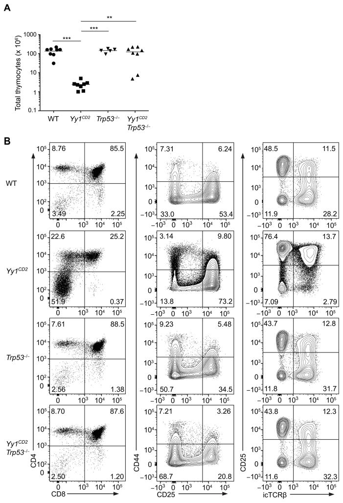
Figure 8. Absence of p53 rescues the developmental defect in YY1CD2 mice.
(A) Number of total thymocytes in Yy1f/f (WT), Yy1f/f CD2-Cre (Yy1CD2), Trp53−/− and Yy1f/f CD2-Cre Trp53−/− (Yy1CD2 Trp53−/−) mice. Each data point represents an individual mouse and horizontal lines indicate the mean. Statistical significance was evaluated by one-way ANOVA with Tukey’s multiple-comparison test. (B) Staining of CD4 and CD8 in total thymocytes (left column), of CD44 and CD25 in DN thymocytes (CD4−CD8−Lin−) (middle column), and of CD25 and icTCRβ in DN thymocytes (CD4−CD8−Lin−) (right column) are shown. The results are representative of two independent experiments. **P < 0.01, ***P < 0.001.

