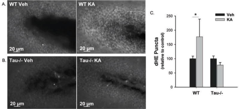
Figure 2. Tau facilitates ROS production in response to excitotoxic insult in vivo.

WT and tau−/− mice received 1mg/kg dHE (i.p.) prior to 25mg/kg KA (i.p.). One hour after KA treatment, dHE fluorescence was detected in the hippocampi of the mice as an indicator of KA-induced excitotoxic ROS production. (A) and (B) show representative 40× images of dHE fluorescence in the dentate gyri of WT and tau−/− mice, respectively, injected with vehicle (left) or 25mg/kg KA (right). The number of dHE puncta was quantified from the entire hippocampus (CA1, CA2, CA3, and DG) and data are presented as the percent change from the vehicle treated group for each genotype averaged over all animals +/− SEM (C). (* indicates P<0.05; WT Veh, n = 8; WT KA, n = 10; Tau−/− Veh, n = 6; Tau−/− KA, n = 6).
