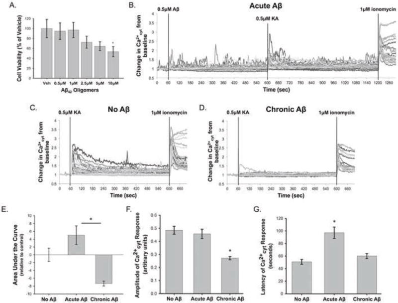
Figure 4. Aβ42 has a biphasic effect on KA-induced Ca2+ influx in primary rat cortical neuron cultures.

Cell viability assays were performed on rat primary cortical neurons using Calcein AM to investigate the toxicity of Aβ42 oligomers at a range of concentrations (0.5μM, 1μM, 2.5μM, 5μM, and 10μM) after a 48 hour treatment (A). Viability data are normalized to the vehicle treated control and are presented as mean +/− SEM (n = 4, * indicates P<0.05). Rat primary cortical neurons were loaded with Fura2-PE3 and were treated with “Acute Aβ” (0.5μM Aβ42 for 10 minutes) (B), “No Aβ” (C), or “Chronic Aβ” (0.5μM Aβ42 for 24 hours) (D) prior to 0.5μM KA treatment. (B–D) show representative Ca2+cyt traces from individual experiments in each condition. Chronic Aβ42 decreased the average area under the KA-treatment portion of the Ca2+cyt curve, while Acute Aβ42 had the opposite effect (E). Chronic Aβ42 treatment also significantly reduced the amplitude of KA-induced Ca2+ influx (F). Acute Aβ42 increased the latency of the Ca2+cyt response from control (G). Data in (D–F) are presented as mean +/− SEM from 12 to 32 separate experiments (* indicates P<0.05).
