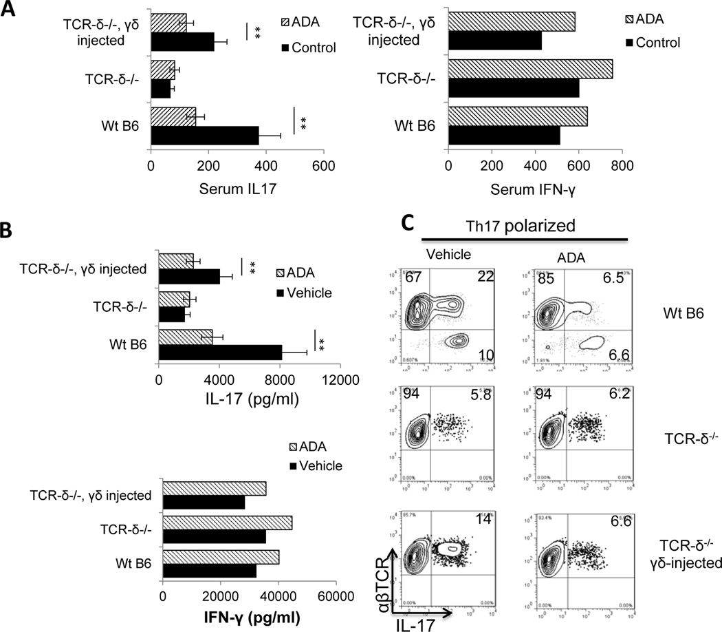
Fig. 4. The effect of ADA is γδ T cell-dependent.
One group of B6 mice and two groups of TCR-δ−/− mice (n=6 for each) were set up and mice in one of the TCR-δ−/− groups were injected with γδ T cells from immunized B6 mice (2 × 10⁶/mouse) immediately before immunization. All groups were then immunized with IRBP1–20/CFA, and ADA or PBS was injected i.p. on day 8 post-immunization.
(A) On day 13-post immunization, serum IL-17 levels (left panel) and IFN-γ levels (right panels) were measured by ELISA
(B) T cells isolated from each group of mice on day 13 post-immunization were stimulated in vitro with immunizing peptide and APCs under Th1-polarizing conditions (upper panel) or Th17-polarizing conditions (lower panel) and the 48 h culture supernatants assessed for IL-17 and IFN-γ by ELISA.
(C) The percentage of IL-17+ cells among the proliferating T cells was assessed after 5 days’ in vitro stimulation of CD3+ T cells taken on day 13 post-immunization with the immunizing peptide and APCs under Th17-polarizing conditions. Data are from a single experiment, representative of three independent experiments. In A and B, **p < 0.05.

