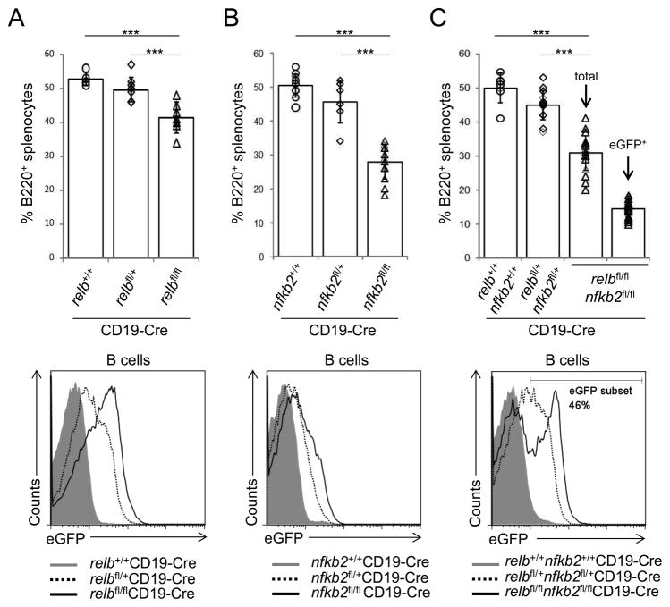
Figure 2. Reduced fractions of splenic B-cells in the absence of alternative NF-κB subunits.
(A–C, top) The fractions of splenic B-cells from relbfl/flCD19-Cre, nfkb2fl/flCD19-Cre or relbfl/flnfkb2fl/flCD19-Cre mice and the corresponding heterozygous and CD19-Cre control mice were determined by flow cytometry (for absolute B-cell numbers, see Supplementary Fig. 2). Each symbol represents a mouse. Data are shown as mean ± standard deviation. Statistical significance was determined by one-way ANOVA (***, P<0.001). (A–C, bottom) Flow cytometry of eGFP expression in splenic B-cells of the indicated genotypes. The number above the gate indicates the percentage of eGFP+ B-cells among B220+ B cells of relbfl/flnfkb2fl/flCD19-Cre mice.

