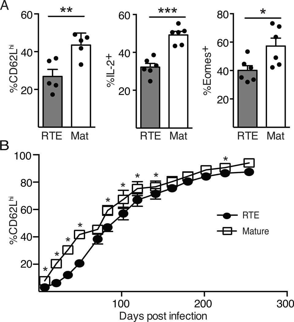
Figure 2. Decreased Tcm generation by RTE-derived memory T cells.
5x10³ each of OT-I TCR Tg RTEs and MN T cells were co-transferred into congenic hosts that were infected the following day with Lm.OVA. A) Splenocytes were analyzed on d60 post-infection. Shown are the percent of RTE and mature CD8+ T cells expressing CD62L and Eomes directly ex vivo and IL-2 production after peptide restimulation. Data are presented as mean ± SEM and are compiled from 2–4 independent experiments. B) Blood was analyzed at the same time points as in Figure 1A to determine the percent of RTE-derived and mature T cells that were CD62Lhi. Data are presented as mean ± SEM and are compiled from 4 independent transfers; n=6–21. * p values vary from <0.05 to < 0.0001 by paired Student’s t test.

