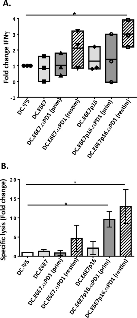
Figure 4. Dendritic cell co-transduced with Ad.E6E7p16 and Ad.αPD1 stimulate greater anti-tumor CD8+ T cell responses mainly when Ad.αPD1 is added at the re-stimulation phase of the in vitro stimulation.
A) CD8 T cells were stimulated for 24 days (two rounds of stimulation) using autologous DC infected with Ad.Ψ5 (DC.Ψ5), Ad.E6E7 (DC.E6E7) or Ad.E6E7p16 (DC.E6E7p16) alone or co-infected with both Ad.E6E7 and Ad.αPD1 (DC.E6E7.αPD1(prim)) or both Ad.E6E7p16 and Ad.aPD1 (DC.E6E7p16.αPD1(prim)). In some cases Ad.αPD1 was only added at the re-stimulation phase on the newly adenovirus-infected DC (DC.E6E7.αPD1(restim) and DC.E6E7p16.αPD1(restim)). Responder CD8 T cells were assessed using IFNγ-ELISPOT assays (A) and 51Cr release killing assay (B) for their functional reactivity on day 24 of in vitro stimulation. IFNγ responses and specific lysis are shown as fold change over DC.Ψ5. *P<.05

