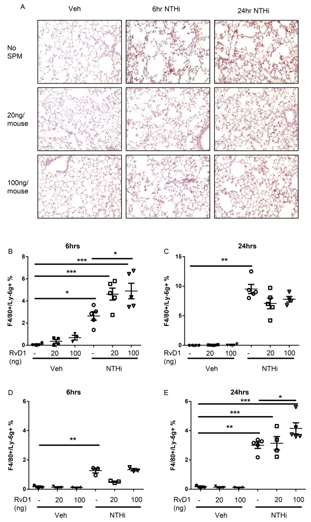
Fig. 3. AT-RvD1 decreases neutrophil influx and promotes enhanced efferocytosis.
Histological sections were taken from AT-RvD1 (20 or 100ng/mouse, OA) and/or NTHi (1 × 10⁶ CFUs, OA) inoculated mice at 6hrs and 24hrs and stained for neutrophils (brown stain) and counterstained with hemotoxylin (A). Efferocytosis from bronchoalveolar lavage (B-C) and collagenase digestion (D-E) at 6hrs (B, D) and 24hrs (C, E) were also evaluated by flow cytometry. F4/80+/Ly6G+ cells represent macrophages with ingested neutrophils. Statistical significance was determined by two-way ANOVA (overall p<0.0001 NTHi [all panels], p=0.0178 AT-RvD1 [B]) with Bonferroni’s posttest for multiple comparisons (*p<0.05, **p<0.01), n=5-8 mice/group.

