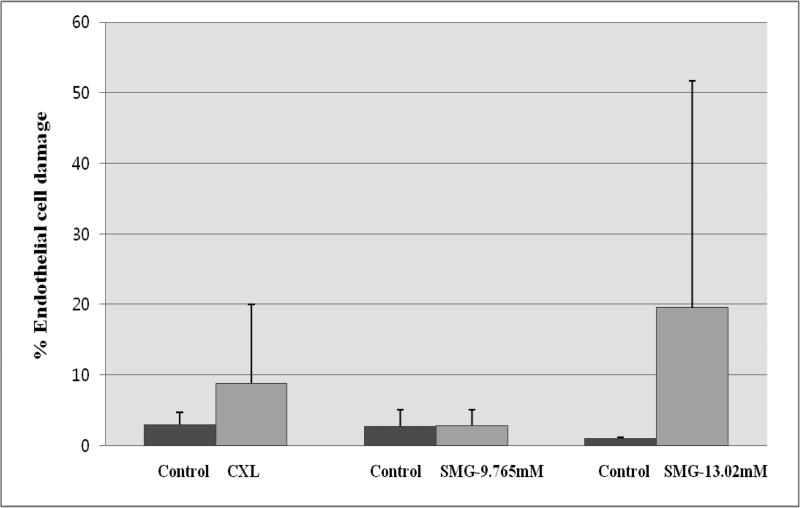
Figure 2.
Endothelial damage from CXL vs. TXL using SMG. Cells were imaged using a confocal fluorescence microscope following live/dead staining using calcein AM (red) and ethidium homodimer (green) [488ex/500-550em]. The method for counting was as described in Methods and Materials. Each condition has its own paired control. All three sets of controls exhibited low levels of dead stained endothelial cells (<3%). SMG at 1/4 max (9.765mM0 was found to induce the least amount of endothelial damage (<3%). 1/3 max SMG was the most toxic (~19%) but a significant sample variability was noted (~30%). CXL showed intermediate endothelial damage at ~8%.

