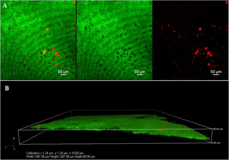
Figure 5.
A. Higher magnification (20×) confocal micrographs of endothelium using calcein –AM (green, live) and ethidium homodimer-1 (red, dead) – stained corneas from UV-CXL using 20X/NA0.75 Plan Apo Nikon objective. B. Tangential sequential image selection for 3-D reconstruction of image A. was carried out. The purpose of this image analysis maneuver was to confirm that the origin of the dead cells was indeed coming from the endothelial monolayer. This was confirmed.

