Figure 1.
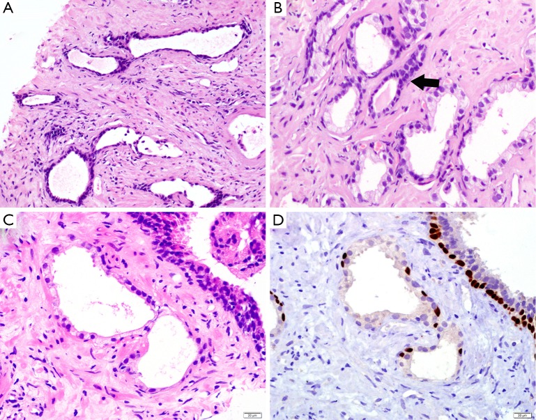
Prostatic atrophy. (A) The glands with simple atrophy show scant cytoplasm and dilated lumens; (B) the cells of post-atrophic hyperplasia (black arrow) have scant cytoplasm and crowded, hyperchromatic nuclei; (C) the cells with partial atrophy lost apical cytoplasm but preserve lateral cytoplasm; (D) the glands of partial atrophy express p63 IHC marker in a discontinuous pattern. (H&E: A, 200×; B&C, 400×. IHC: D, 400×). IHC, immunohistochemistry.

