Figure 1.
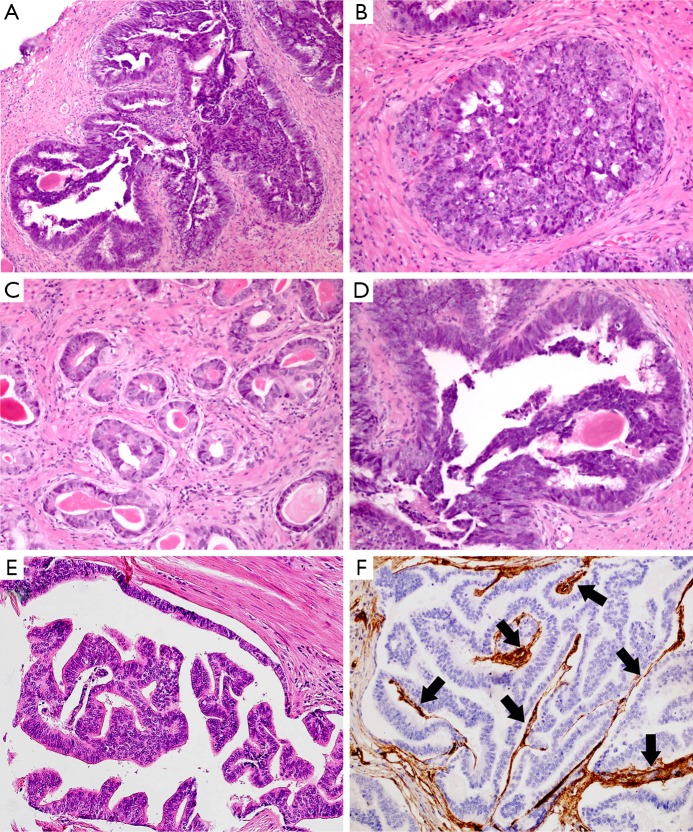
PDA exhibits papillary (A), cribriform (B), and glandular (C) growth pattern. The tumor cells are pseudostratified columnar epithelium with amphophilic cytoplasm, elongated nuclei and prominent large nucleoli (D). This entity is characterized by the presence of tall, pseudostratified, columnar cells with abundant cytoplasm arranged in papillary pattern (E). The central fibrovascular cores in the papillae are highlighted by CD34 immunohistochemical staining (F, black arrows: blood vessels). (H&E: A, B, C&E, 200×, D, 400×; IHC, F, 200×).

