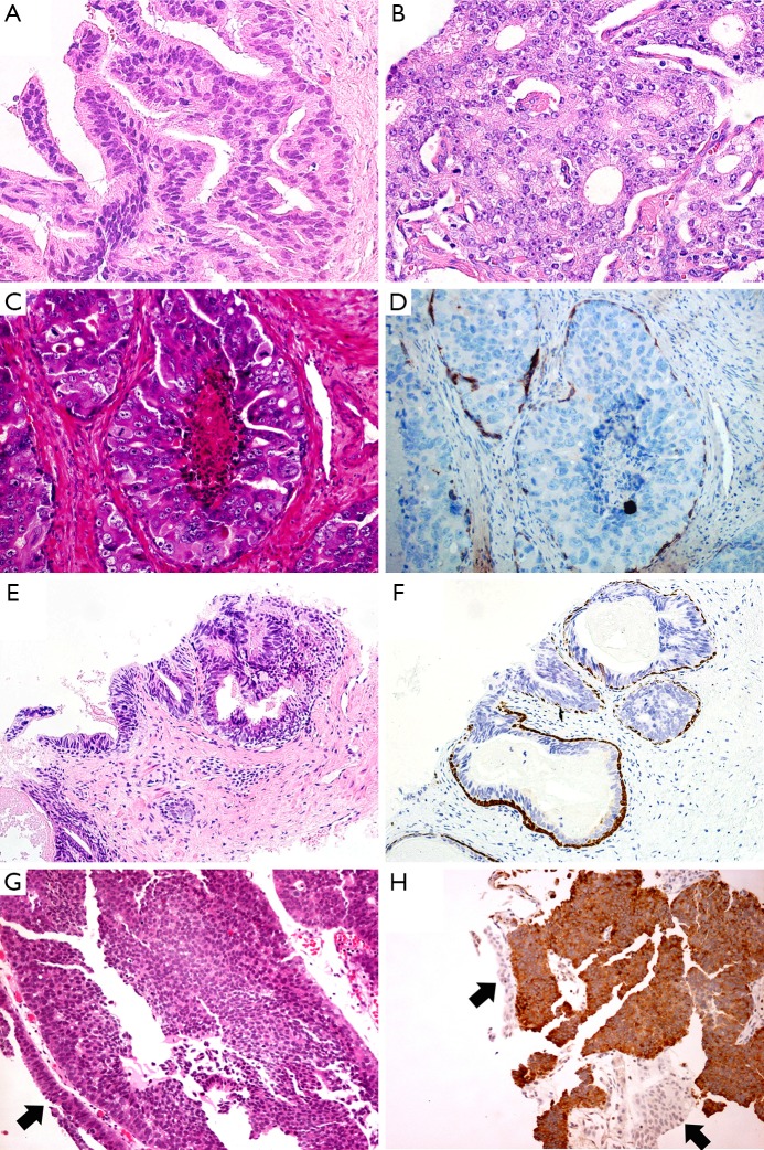
Figure 2.
The cribriform pattern of PDA exhibits the irregular slit-like lumens with tall pseudostratified columnar neoplastic cells (A). The cribriform characteristic of PAA is punched-out round lumens with cuboidal tumor cells (B). IDAC-P exhibits comedo-necrosis in the lumen. The tumor cells are cuboidal, with very large and atypical round or oval nuclei with prominent nucleoli (C). The basal cells present at the periphery in a patchy pattern (D). The columnar tumor cells in HGPIN mimic PDA (E). Basal cells outlined by HMWCK and P63 suggest the diagnosis of HGPIN (F). One case of PDA was misdiagnosed as urothelial carcinoma in core needle biopsy specimen. The tumor cells infiltrate into prostatic urethra (G). Expression of PSA favors the diagnosis of PDA (H). (H&E: A&B, 400×, C, D&G, 200×; IHC: D, F&H, 200×; black arrows: normal urothelium).

