Figure 4.

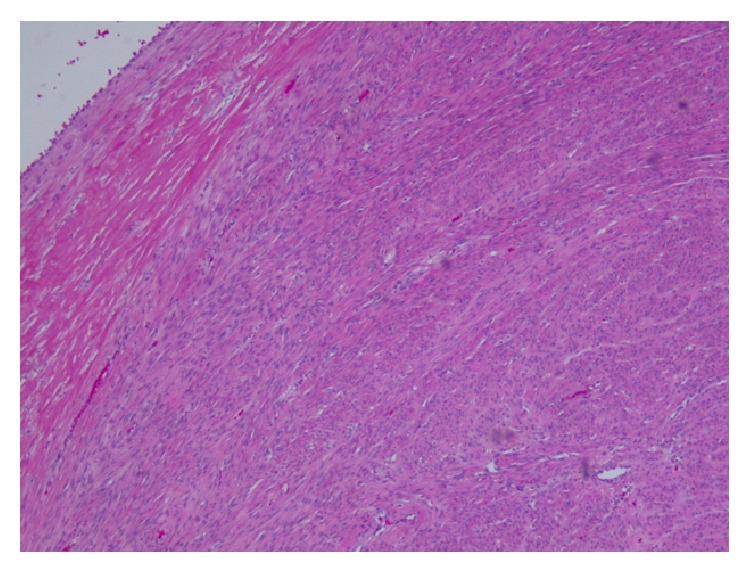
Section from one of peritoneal nodules showing proliferation of spindle cells without atypia, mitosis, or necrosis (Hematoxylin and Eosin stain, 100x Magnification).

Section from one of peritoneal nodules showing proliferation of spindle cells without atypia, mitosis, or necrosis (Hematoxylin and Eosin stain, 100x Magnification).