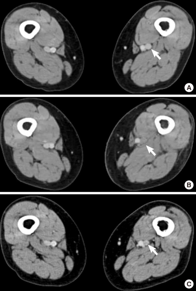
Fig. 2.

Serial MDCT venography images showing the natural course of postoperatively developed proximal DVT following unilateral UKA in a 71-year old female patient. Normal venous flows are observed at both proximal thighs preoperatively. Arrow indicates the left poplieal vein (A). Mild engorgement of the popliteal vein (arrow) is noted in the left proximal thigh at postoperative 1-week (B). At postoperative 6-month, the DVT lesion is completely regressed without thrombolytic treatment. Arrow indicates the left popliteal vein (C).
