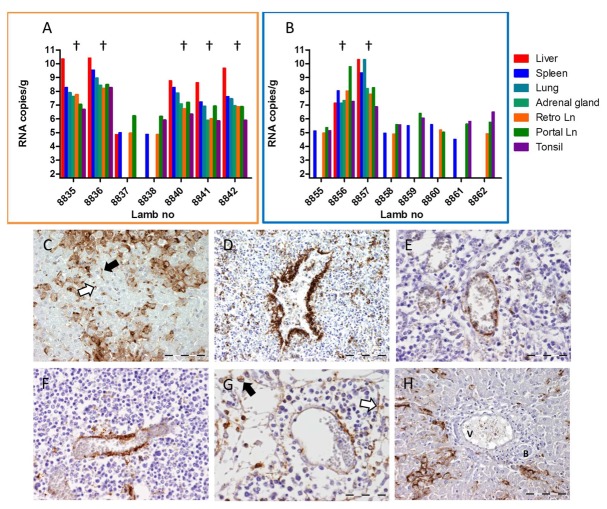
FIGURE 3.
Detection of viral RNA and viral antigen in organ samples collected from animals of Experiment B. Detection of viral RNA by qRT-PCR in organ samples collected from contact-exposed and subsequently challenged lambs (orange frame, A) and of infected control lambs (blue frame, B). Lambs that succumbed to the infection are marked (†). (C–H) Immunohistochemical staining of organ samples collected from lamb 8836. (C) Detail of liver acinus with strong immunostaining of hepatocytes. Also note the accumulation of RVFV antigen in Kupffer cells (black arrow) and circulating macrophages (white arrow) within the liver sinusoids. (D–G) Positive staining for RVFV antigen in the endothelial cells of venules and veins in (D) spleen; (E) tonsil; (F) paracortex of the retropharyngeal lymph node; and (G) medulla of the portal lymph node. Also note the positive staining in the circulating macrophages (black arrow) and littoral cells (white arrow) in the medullary sinuses of the lymph node. (H) Portal area of the liver. Note the heavy staining for RVFV antigen in the hepatocytes and the absence of staining of the endothelial layer of the Vena porta (V). B = bile duct. Bar = 100 μm (C,D,H) and 50 μm (E,F,G).

