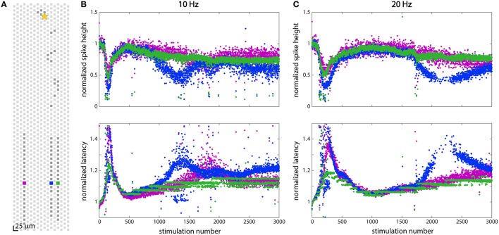
Figure 6.
Stimulation at 10 Hz and 20 Hz showed different effects in two axonal branches. (A) Position of electrodes in three channels from which spikes were recorded. (B) Responses to stimulation at 10 Hz at the three electrodes shown in (A). (C) Responses to stimulation at 20 Hz at the three electrodes shown in (A). At both 10 and 20 Hz, all three axonal branches showed slightly different responses to stimulation, although the blue branch showed the most dramatic effects. All three branches display the same adaptation characteristics following 150 (at 10 Hz) or 200 (at 20 Hz) stimulations, but additional changes in the wave forms are seen in some branches while not in others.

