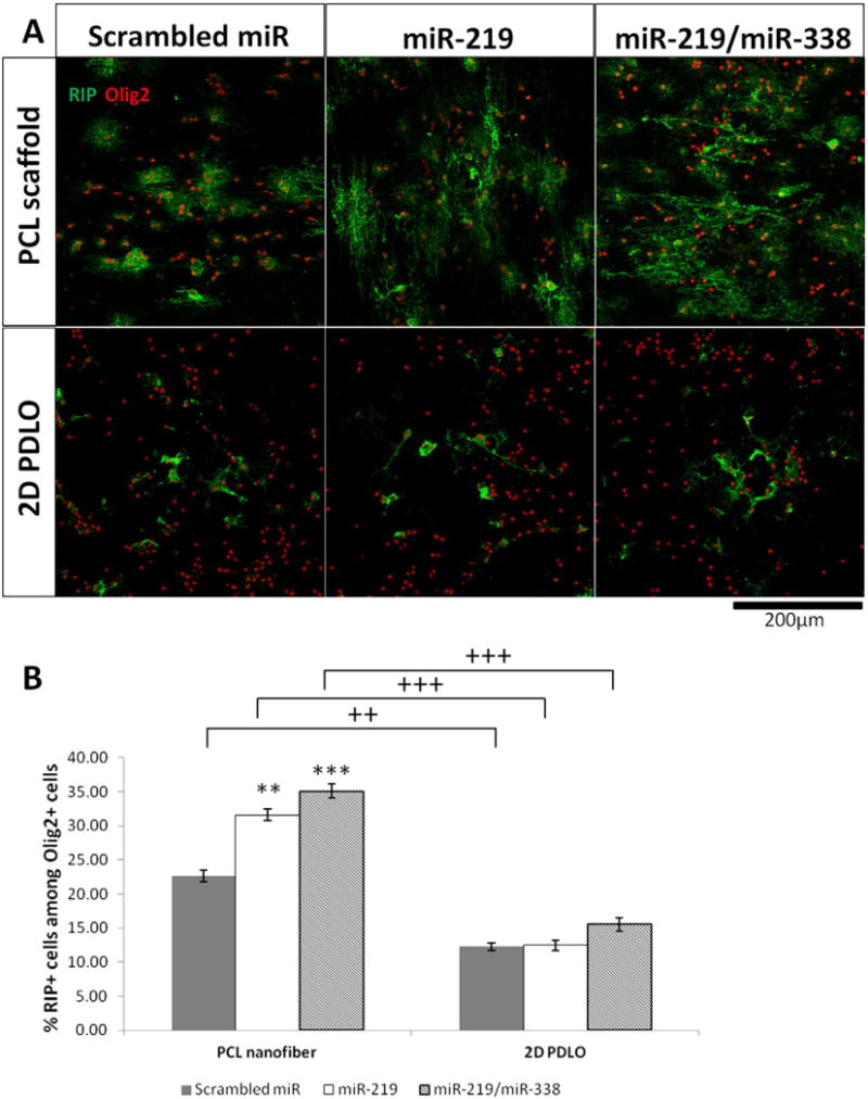
Fig. 4.

Immunofluorescent staining (day 4) of immature OL marker RIP, indicating enhanced differentiation on PCL scaffold. (A) Representative fluorescent images and (B) Quantification of RIP+ cells among Olig2+ oligodendrocyte lineage cells. **p < 0.01 and ***p < 0.001 versus respective scrambled miR group (ANOVA, mean ± S.E.M., N = 3). ++p < 0.01 and +++p < 0.001 between two groups with same miR treatment (Student’s t-test).
