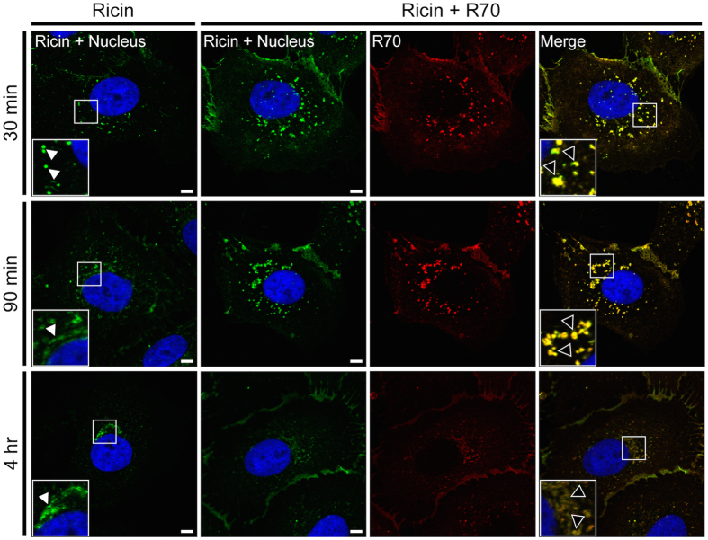
Figure 1. R70 is internalized in complex with ricin and delays toxin accumulation in TGN.
Vero cells, grown on glass coverslips, were cooled to 4 °C and incubated for 30 min with ricin-FITC. The cells were then washed, treated (or not) with R70 for an additional 30 min at 4 °C and then shifted to 37 °C. At the indicated time points (30 min, 90 min and 4 hr) the cells were fixed, stained with DyLight® 549-labeled secondary Ab and imaged by confocal microscopy. In the images, ricin appears green, R70 is red, a merge between ricin and R70 is yellow, and the cell nucleus is blue. Insets in the right and left hand columns highlight the subcellular localization of ricin (white arrowheads) and ricin-R70 complexes (black arrowheads). Images are representative of at least 5 independent experiments. Scale bar, 5 μm.

