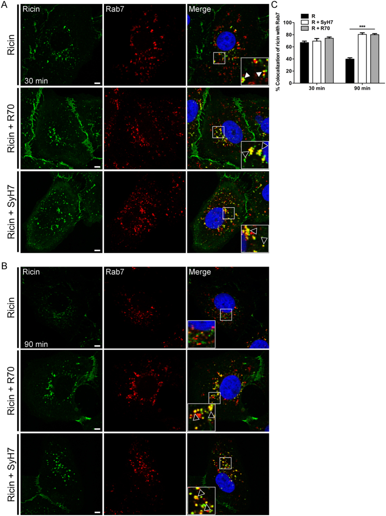
Figure 5. R70 and SyH7 delay ricin egress from Rab-7 vesicles.
Vero cells treated at 4 °C with ricin-FITC (top panels), ricin-FITC and R70 (middle panels), or ricin-FITC and SyH7 (bottom panels), as described in the Materials and Methods, were shifted to 37 °C for (A) 30 min, or (B) 90 min before being fixed and stained for Rab7 (red). Insets (right column) indicate minimal visual colocalization between ricin and Rab7 in the absence of R70 and SyH7, but notable colocalization in the presence of R70 and SyH7 (arrowheads) at both time points. Scale bars, 5 μm. Images are representative of at least three independent experiments. (C) The frequency of ricin colocalization with Rab7 at the 30 and 90 min time points were quantitated with ImageJ, as described in the Materials and Methods. Each bar represents the average of approximately 20 cells (with SEM) of 3 individual experiments. ***p < 0.001, determined using a one-way ANOVA with Tukey’s post-test.

