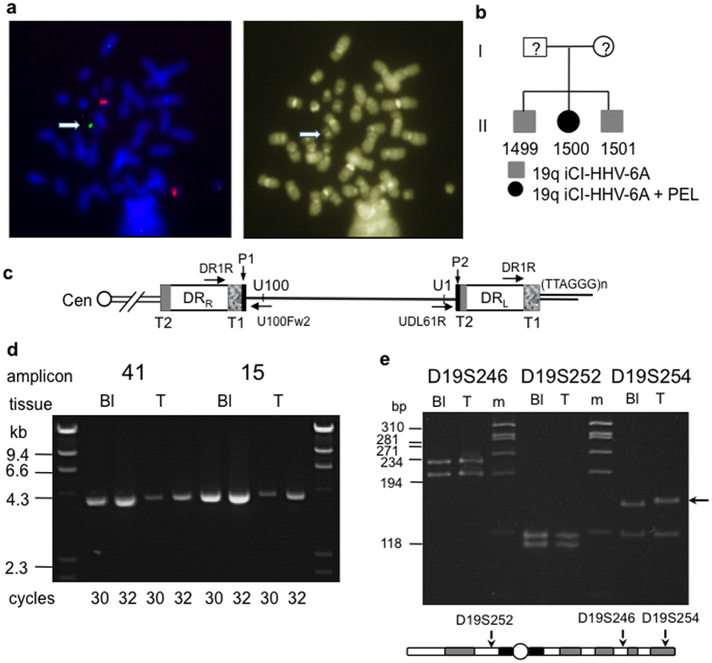
Figure 1. iciHHV-6A at 19q in three siblings and loss from the HHV-8-unrelated PEL-like lymphoma.
(a) FISH on metaphase chromosomes from 1500-Bl (blood). The green labeled HHV-6 probe showed HHV-6 sequence at the telomere of chromosome 19q, a probe for centromere 20 (red signals) served as a control (b). Pedigree of family with iciHHV-6A at 19q. (c) Organisation of iciHHV-6A in the 19q telomere. Diagram shows location of priming sites for DR1R, U100Fw2 and UDL61R. T1 and T2 are the imperfect and perfect arrays of viral encoded (TTAGGG)n repeats respectively. P1 and P2 show the locations of the PAC1 and PAC2 sequences retained in the integrated viral genome. (d) Examples of semi-quantitative analysis of two HHV-6A amplicons (41 and 15) in 1500-Bl and 1500-T (pleural fluid containing HHV-8-unrelated PEL-like lymphoma cells). (e) 1500 was heterozygous at three STRs on chromosome 19 (D19S252, D19S246, D19A254) in blood DNA (Bl) and heterozygosity is retained in the lymphoma (T). Black arrow shows the mutated D19S254 allele.

