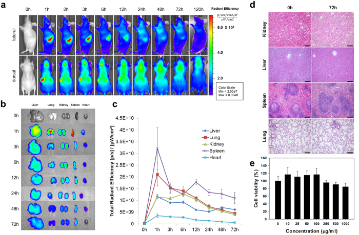
Figure 2. Biodistribution and Toxicity Assay of MMR-Cy5.5.
(a) In vivo fluorescence whole body imaging to evaluate the distribution of MMR-Cy5.5 in C57BL/6 nude mice at different time points after injection. (b) Ex vivo fluorescence imaging of the harvested liver, lung, kidney, spleen, and heart from MMR-Cy5.5-injected wild type C57BL/6 mice analyzed at scheduled time-points. (c) The time dependent changes of fluorescence intensities of isolated organs from ex vivo images. (d) H&E stained images of the kidney, liver, spleen, and lung from MMR-Cy5.5-injected mice for toxicity analysis. No histopathological changes were observed at 72 h post-injection. Scale bar, 200 μm. (e) Cellular toxicity analysis shows that 85–95% of the cells are still viable after incubation with various concentrations of MMR-Cy5.5 up to 1000 μg/mL for 24 h.

