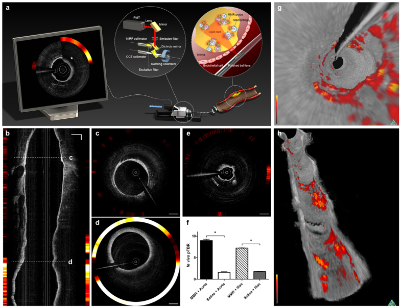
Figure 4. In Vivo Imaging of Inflamed High-risk Plaques in Coronary-sized Vessels using MMR-Cy7 and the OCT-NIRF Catheter-Based Imaging System.
(a) Schematic diagram of the high-speed dual-modal OCT-NIRF catheter-based imaging system. The rotary junction in the center rotates at 100 rps. The ball lens simultaneously collects the OCT and the NIRF signals, which propagate through the double-clad fiber (DCF). The two different signals are separated with the dichroic mirror and are rendered through a custom-built software showing the final OCT-NIRF image presented on the monitor (asterisk), which demonstrates enhanced NIRF signals existing in the macrophage-abundant plaque lesion identified by OCT. (b) A longitudinal OCT-NIRF image and (c) the corresponding cross-sectional images of a normal lesion, (d) an advanced and (e) early atherosclerotic plaque lesion. Compared to the normal-looking segment, the NIRF signals emits more prominently from the macrophage-abundant atherosclerotic plaque. (f) The in vivo pTBR is significantly higher in the MMR-Cy7 injected rabbits compared to the saline-injected control group (*p < 0.05) regardless of the vessel site. (g,h) Three-dimensional OCT-NIRF rendered images. (g) A fly-through and (h) a longitudinal cutaway view enables the identification of both NIRF signals and luminal morphology. Scale bars, 1 mm.

