
Figure 2. Xyloglucan degradation patterns by purified cellulosomes, Xgh74A, Cel44O, or Cel9X.
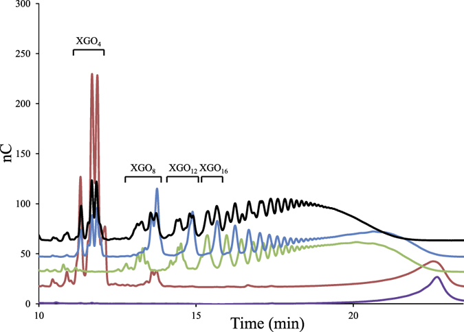
The samples were analyzed by HPAEC-PAD. Xyloglucan at 3.5 g/L was incubated for 3 h at 37 °C with no enzyme (purple), with 6 mg/L (approx. 10 nM) of purified cellulosomes (black), with 2.5 nM of Xgh74A (red), with 2.5 nM of Cel44O (green), or with 2.5 nM of Cel9X (blue). “XGO4”, “XGO8”… “XGO16” refer to xyloglucan oligosaccharides displaying 4, 8…16 glucosyl residues backbone, respectively.