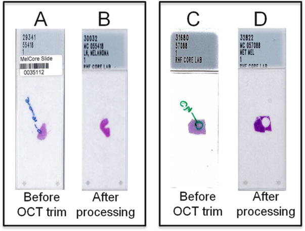
Fig. 1.

H&E-based “trimming” of OCT specimens. Examples of H&E-stained OCT tumor sections before (A and C) and following (B and D) trimming of OCT blocks to maximize tumor cell nuclei and to remove areas of necrosis. Note that circled areas (A and C) were identified for trimming
