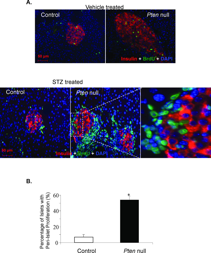
Figure 2. Peri-islet proliferating cells induced by STZ treatment do not express insulin.
(A) Control and Pten null mice were treated with either vehicle sodium citrate (top row) or STZ to induce β-cell injury (bottom row) and the pancreas was analyzed with the endocrine β-cell marker insulin (red), proliferating cell marker BrdU (green) and nuclear cell marker DAPI (blue). Top row: The left panel is the control pancreas. The right panel is the Pten null pancreas. Original magnification, 20×. Bottom row: The left panel is the control islet. The middle panel is the Pten null islets. Original magnification, 20×. The right panel is a higher magnification of the area within the dashed square. (B) Graph summarizing the ratio of the number of islets with increased per-islet proliferation compared to the number of islets with little to no peri-islet proliferation (n=4). The bars represent the average percentage of peri-islet proliferation for the controls and mutants. The error bars represent the SEM for all animals analyzed for that group. *p≤0.05 is considered to be statistical significant. Bar, 50µM.

