Figure 3.
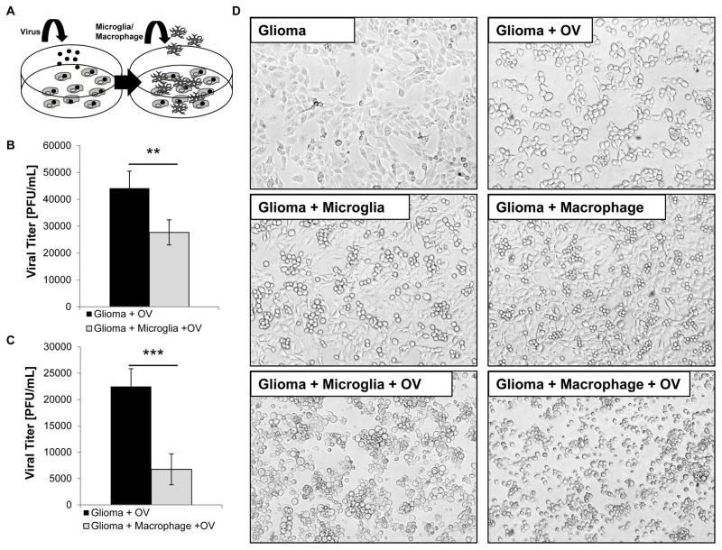
Microglia and macrophages reduce virus replication in tumor cells in vitro. A. Schematic of microglia/macrophage and tumor cell co-cultures. Tumor cells [oval-shaped cells] were infected with oHSV [rHSVQ1] [black dots] at a MOI of 2. Unbound virus was washed away and microglia or macrophages [branched cells] were overlaid on the infected glioma cells. The cells were cultured for 12 hours and the viral titers were determined by a standard plaque formation assay. B–C Viral titers of glioma cells infected alone or cultured with BV2 microglia [n=4/group] [B.] or RAW264.7 macrophages [n=4/group] [C.]. Data shown is mean virus titer ± SD. D. Representative images of glioma cells cultured with microglia and macrophages with and without oHSV infection 12 hours post infection [n= 4/group].

