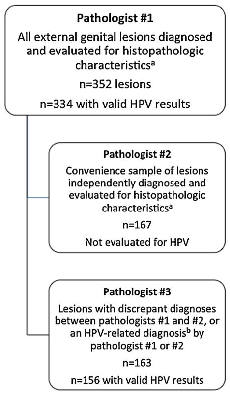
Figure 2.
Overview of specimen evaluation. aHistopathologic characteristics included rounded papillomatosis (Fig. 1A), parakeratosis (Fig. 1B), hypergranulosis (Fig. 1C), dilated vessels (Fig. 1D), koilocytes and binucleation (Fig. 1E), horn cysts, hyperpigmentation, and dysplasia/atypia. bHPV-related diagnoses included condyloma, penile intraepithelial neoplasia I, and penile intraepithelial neoplasia II/III. Note: The lesions evaluated by Pathologist #2 and 3 are not mutually exclusive. Only 155 lesions were utilized for the three-way concordance analysis.

