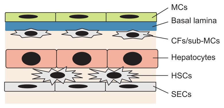
Fig. 3.
Structure of the mesothelium in mouse liver tissue. Mesothelial cells (MCs) line up on the liver surface and form a single epithelial cell layer. The basal lamina separates the MCs from the underlying capsular fibroblasts (CFs)/sub-mesothelial cells (sub-MCs). Mouse livers show a single stratum of CFs beneath the MCs. Hepatic stellate cells (HSCs) reside in the space of Disse between hepatocytes and sinusoidal endothelial cells (SECs).

