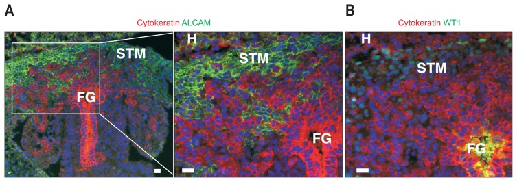
Fig. 4.
Mouse liver development at embryonic day 10. Foregut endoderm (FG) differentiates into hepatoblasts that are positive for immunohistochemical staining of cytokeratin (red). Cytokeratin+ hepatoblasts invade the surrounding septum transversum mesenchyme (STM) and form liver buds. The STM expresses mesothelial cell markers, such as activated leukocyte cell adhesion molecule (A, green) and Wilms tumor 1 homolog (WT1) (B). WT1+ mesenchymal cells also differentiate into epicardial cells of the developing heart (H). Scale bar, 20 μm.

