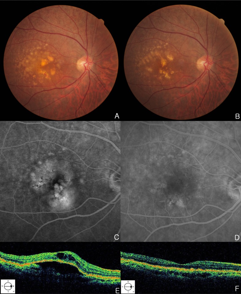
Fig. (1).
(A, C, E) Fundus photographs (A), FFA (C) and OCT (E) images of patient at the first examination. Fundus photographs, a centrally located DPED are seen with diffuse coalesced soft drusen at right eye. Hyperfluorescence of FFA and hypo-reflectivity of OCT correspond to CNV with a DPED. (B, D, F) Fundus photographs (B), FFA (D) and OCT (F) images of patient after ranibizumab injections. After 5 intravitreal ranibizumab injections, six months later, DPED at foveal area and drusen located superotemporal to macula are diminished over time. The late-phase fluorescein study shows regression of CNV.

