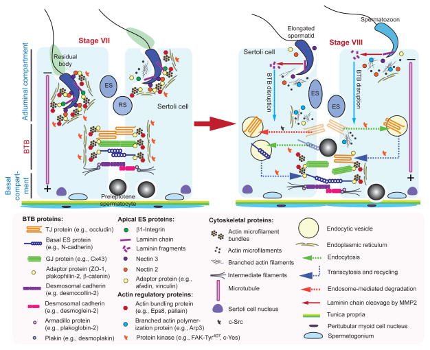
Figure 2. A schematic drawing illustrating the molecular mechanism by which preleptotene spermatocytes are transported across BTB at stage VIII of the epithelial cycle.
The cross-section of a stage VII seminiferous tubule on the left panel illustrates the intact BTB is conferred by adhesion protein complexes of the actin-based TJ, basal ES and GJ, as well as the intermediate filament-based desmosome. The apical ES is also intact which is conferred by the corresponding adhesion complexes of nectin-afadin and β1-integrin-laminins. Preleptotene spermatocytes transformed from type B spermatogonia are first detected at stage VII, and are being transported across the BTB, when apical ES also undergoes degeneration to prepare for spermiation as noted in a stage VIII tubule shown on the right panel. Biologically active laminin fragments (e.g., F5-peptide) generated by MMP-2-induced proteolytic cleavage induce BTB restructuring in which TJ (e.g., occludin), basal ES (e.g., N-cadherin), GJ (e.g., connexin 43), and desmosome (e.g., desmoglein-2) proteins undergo endocytic vesicle-mediated endocytosis, transcytosis and recycling. In this way, the “old” BTB proteins located above the preleptotene spermatocytes can be recycled to assemble a “new” BTB behind these germ cells to facilitate the transport of spermatocytes at the site, prior to the degeneration of the “old” BTB. As such, the events of spermiation and BTB remodeling which allow the release of sperm and the transport of preleptotene spermatocytes, respectively, occur at the opposite ends of the epithelium and take place simultaneously at stage VIII of the cycle.

