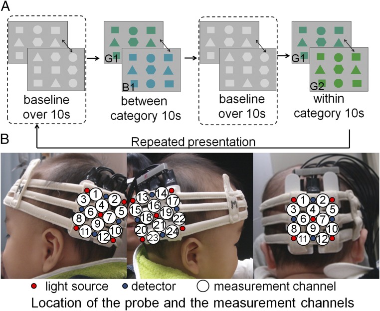
Fig. 1.
Experimental procedure and location of the NIRS probes. (A) In each trial, the baseline phase consisted of figures changing in shape, which had a duration of at least 10 s. The test phase consisted of color changes between categories (G1/B1) or within a category (G1/G2), which had a duration of 10 s. The presentation order of the between- and within-category phases was counterbalanced across infants. (B) Location of the NIRS probes and the measurement channels in the probe system for infants. The probe holders were placed on the left and right OT regions, slightly below T5 and T6 of the international 10–20 system in the main measurement. The probe holder was placed slightly above Oz for the measurement of the occipital region responses. The distance between the emitter and detector probes was set at 2 cm.

