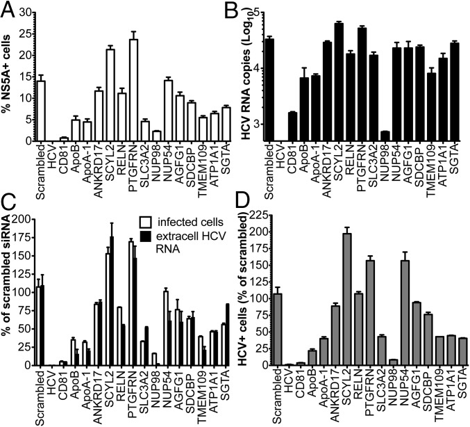
Fig. 5.
Validation of cellular proteins incorporated in HCV virions by RNAi. Huh-7.5 cells were transfected with siRNA smart pools against the indicated molecules and infected 48 h later with Con1/Jc1 at low MOI (0.05). (A and C) Percentage of HCV+ cells was determined 3 d postinfection (dpi) by NS5A staining and FACS analysis (white bars). (B and C) Number of HCV RNA genomes released in the cell media was quantified by RT-quantitative PCR (black bars). (D) Infectivity of HCV particles produced by siRNA-transfected cells was assessed by inoculating equal volumes of cell culture media on naive Huh-7 cells. The number of NS5A+ cells was determined by FACS 3 dpi. In C and D, data are expressed as a percentage of scrambled siRNA.

