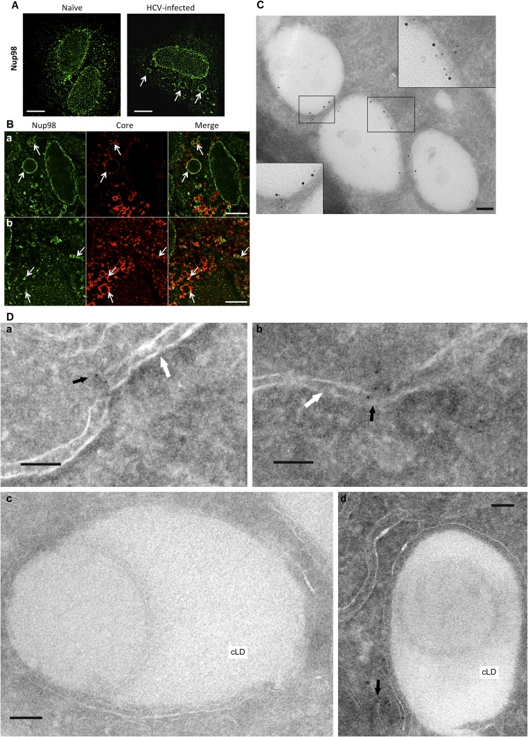
Fig. S5.
Subcellular localization of endogenous Nup98 in Huh-7.5 cells. Naive (A) and HCV-infected (B) cells were stained (6 dpi) with anti-Nup98 (green channel) and anti-HCV Core (red channel) Abs. Immunofluorescence images are shown. (Scale bars, 10 μm.) (A) Unusual staining patterns for Nup98 are observed in infected cells (arrows). (B) Colocalization of Nup98 with Core at these circular structures from two independent fields (a and b). (C) Huh-7.5 cells infected with clone 2 for 4 d were cryosectioned and stained with Abs to HCV Core (12 nm) and Nup98 (6 nm), using the Tokuyasu (9) method. Representative iEM images are shown. (Scale bars, 100 nm.) (D) Subcellular localization of endogenous Nup98 in naive Huh-7.5 by iEM. Cells were cryosectioned and stained with anti-Nup98 Ab (6 nm). Nup98 mostly localized at the nuclear membrane (a and b), whereas no staining on the surface of cLDs was observed (c and d). Representative iEM images are shown. Black arrows show Nup98, and white arrows indicate the nuclear membrane. (Scale bars, 100 nm.)

