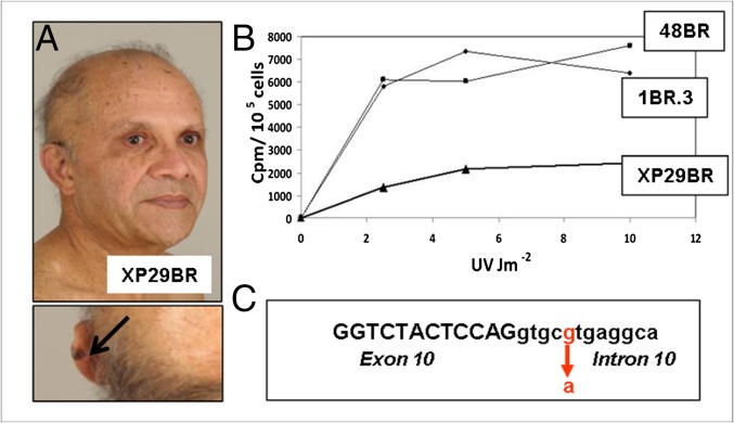
Fig. 2.
XP29BR. (A) Patient XP29BR, with a melanoma on his left ear (arrow). (B) UDS, measured as incorporated 3H-thymidine cpm per 105 cells following UV irradiation of XP29BR with different UVC doses compared with normal fibroblasts 1BR.3 and 48BR. (C) Sequence around the exon 10/intron 10 boundary showing the position of the mutation.

