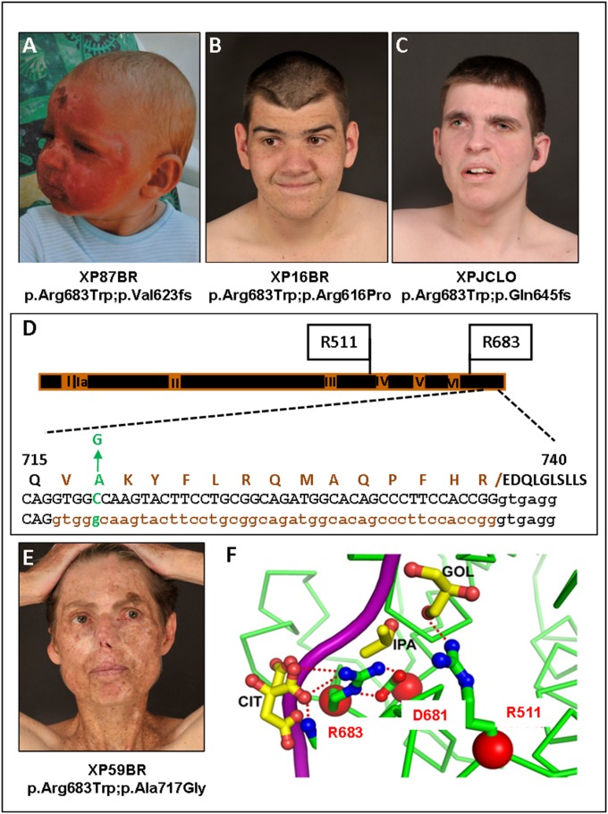
Fig. 3.
XP-D patients. (A) Severe sunburn after minimal sun exposure in XP87BR before XP diagnosis (see ref. 5 for another photograph of this patient). (B) XP16BR and (C) XPJCLO showing minimal pigmentary skin changes due to early diagnosis and excellent UVR protection. (D) Scheme of XPD protein, showing the seven helicase domains, I, Ia, II–VI, and the positions of the mutations in the XPD patients. Below is an expanded view of aa715–740 showing the p.Ala717Gly alteration and the deletion of aa716-730 resulting from the c.C2150 > G mutation. Top line: WT protein sequence with p.Ala717Gly indicated in green and the deleted 15 aa resulting from abnormal splicing of intron 22 indicated in brown; middle and bottom lines: WT and mutant DNA with exon 22 sequence in caps, intron 22 in lowercase, with abnormally spliced-out bases in brown. (E) XP59BR, showing multiple surgical scars as a result of poor UVR protection (see ref. 5 for another photograph of this patient). (F) Crystal structure of XPD around Arg683, showing projected hydrogen bonding with Asp681 and DNA (magenta). (Modified from ref. 33.)

