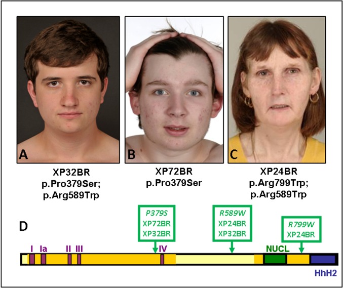
Fig. 4.
XP-F patients. (A) XP32BR. (B) XP72BR. (C) XP24BR. Note the very mild skin changes despite only moderate UVR protection. (D) Scheme of XPF protein, showing the five motifs of the disrupted helicase domains (I, Ia, II, III, and IV), the nuclease domain (NUCL), and the two Helix-hairpin-helix (HhH) domains.

