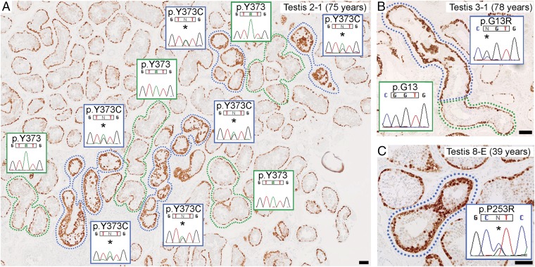
Fig. 1.
Seminiferous tubules strongly immunopositive for MAGEA4 contain pathogenic mutations. (A–C) Thin sections from three FFPE testes showing spermatogonia, marked by MAGEA4 positivity (brown staining), present in a single layer at the periphery of normal tubular cross-sections (green surround or unlabeled). A subset of tubules (immunopositive tubules) display enhanced MAGEA4 staining (blue surround) due to dense clustering of spermatogonia with strong immunoreactivity. Dideoxy-sequencing traces were obtained from non-WGA DNA extracted from microdissected tissue of an adjacent section. (A) Heterozygous FGFR3 c.1118A>G (p.Y373C) mutations (*) are present in immunopositive tubules, but not in neighboring normal tubules. Clusters of mutation-positive tubules likely represent cross-sections of a single convoluted tubule. (B) In a longitudinal section of a tubule showing the transition from normal to strongly immunopositive staining, the heterozygous HRAS c.37C>G (p.G13R) mutation (*) was specific to the immunopositive portion, pinpointing the boundary between nonmutant and mutant cells. (C) Heterozygous FGFR2 c.758C>G (p.P253R) mutation (*) in immunopositive tubule. (Scale bars: 100 µm.)

