Fig. 7.
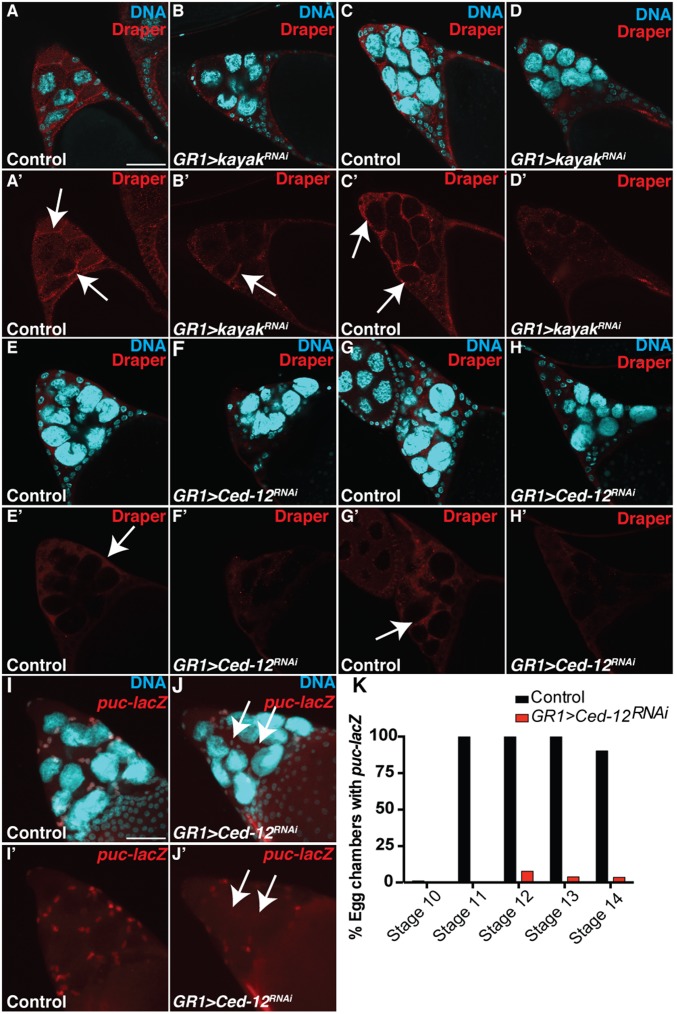
Ced-12 acts upstream of JNK in late oogenesis. (A–D’) Control (PG150-GAL4/+; UAS-mCD8-GFP/+) and kayak knockdown (GR1-GAL4/UAS-kayakRNAi) egg chambers were stained with α-Draper antibody (red) and DAPI (cyan). (Scale bar, 50 μm.) (A–B’) The initial expression of Draper during stage 11 is unaffected. (C–D’) The enrichment of Draper on the FC membranes in kayak knockdown in stage 12 (D and D’) is reduced compared with the control (C and C’). (E–H’) Control (GR1-GAL4/UAS-mCD8-GFP) and Ced-12 knockdown (GR1-GAL4/UAS-Ced-12RNAi) egg chambers were stained with α-Draper antibody (red) and DAPI (cyan). (E–F’) The initial enrichment of Draper on the FC membranes during stage 11 is not apparent in the Ced-12 knockdown. (G–H’) The enrichment of Draper on the FC membranes in the Ced-12 knockdown in stage 12 (H and H’) is absent compared with the control (G and G’). (I–J’) Projection images (34 slices) of stage 11 egg chambers stained with DAPI (cyan) and α–β-Gal (red) as a readout of JNK activity. (I and I’) Most stretch FCs in control puc-lacZ/TM3 egg chambers express β-Gal. (J and J’) puc-lacZ/GR1-GAL4 UAS-Ced-12RNAi egg chamber FCs have reduced β-Gal expression (arrows). (K) Quantification of puc-lacZ in Ced-12 knockdowns (GR1-GAL4 Ced12RNAi/puc-lacZ) compared with the control (puc-lacZ/TM3). Egg chambers were considered to have activated puc-lacZ when >10 FCs were stained with α–β-gal.

