Fig. 1.
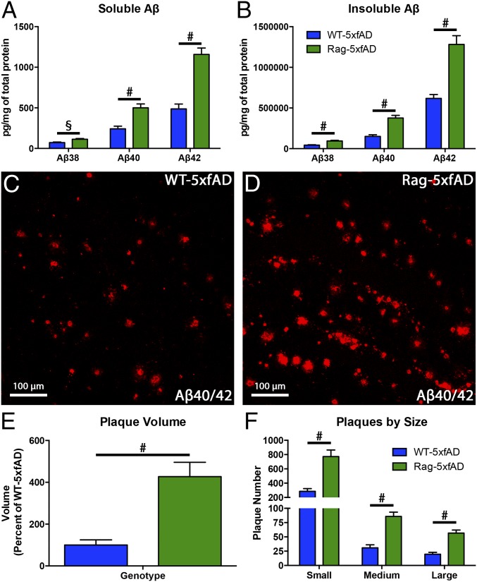
Aβ is significantly elevated in immune-incompetent Rag-5xfAD mice. (A and B) Aβ multiplex ELISA reveals significant elevations in all three Aβ species in both soluble and insoluble brain lysates from Rag-5xfAD vs. WT-5xfAD. Representative confocal images of Aβ40/42-immunoreactive plaques within the dentate gyrus demonstrates a similar robust change in plaque load between WT-5xfAD (C) and Rag-5xfAD mice (D). (E) IMARIS-based 3D quantification of Aβ confirms ELISA and immunohistochemical (IHC) findings, demonstrating a more than fourfold increase in dentate gyrus plaque volume in Rag-5xfAD mice (green) vs. WT-5xfAD mice (blue). (F) Stratification of dentate plaque numbers by size likewise reveals significant elevations in Rag-5xfAD mice. Data are represented as mean ± SEM. ANOVA, P < 0.05, and Fisher’s protected least-significant difference (PLSD) post hoc, §P < 0.01, #P < 0.001; n ≥ 8 mice/group.

