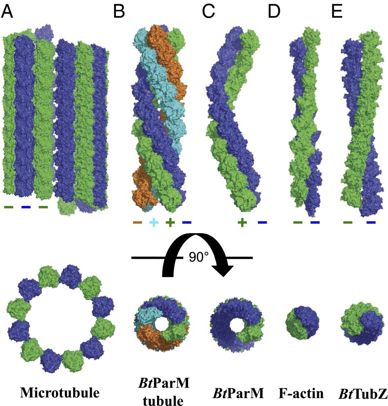
Fig. 5.
Comparison of actin-like and tubulin-like filament structures. (A) Mammalian microtubule. (B) B. thuringiensis actin-like BtParM nanotubule. (C) B. thuringiensis actin-like BtParM filament. (D) Mammalian F-actin. (E) B. thuringiensis tubulin-like TubZ. The filament systems are depicted with 10 protomers in each strand, with the exception of F-actin, which has eight. (Upper) Side views. (Lower) Top views.

