
Figure 2. Location of the volume of interest in the PMC.
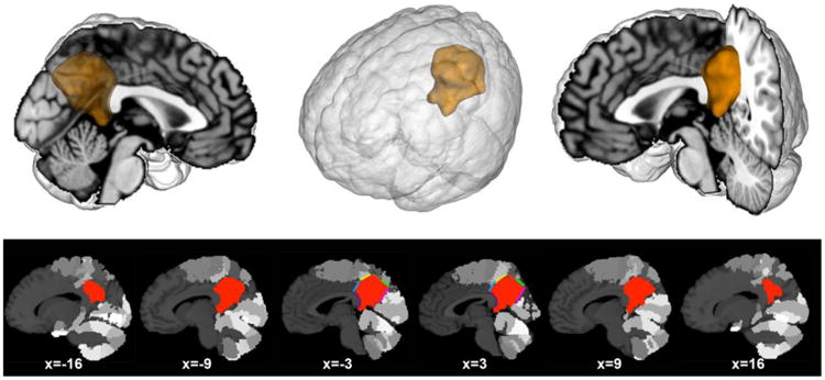
The volume of interest (VOI) was created manually guided by bordering macroanatomical landmarks and cytoarchitectonic areas from the Jülich brain atlas (cf. method section). Our VOI comprised the posterior medial cortex providing the starting point for the present analyses. Upper row: the VOI was rendered on a T1-weighted MNI single-subject template using Mango (multi-image analysis GUI; http://ric.uthscsa.edu/mango/). Lower row: sagittal sections through the VOI based on the same template. Colors indicate anatomical borders that informed the VOI definition: blue (area 23d), yellow (5M), red (7A), green (7P), pink (parietooccipital sulcus), and purple (splenium of corpus callosum). The gray colored clusters of the brain template indicate cytoarchitectonic areas surrounding the VOI. Coordinates in MNI space.