
Fig 2. Waddlia chondrophila replicate within mouse lungs.
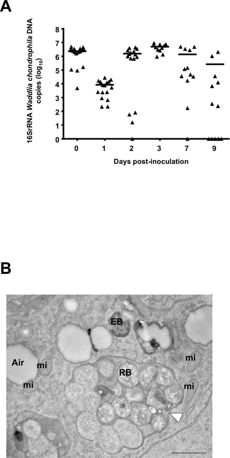
A) Bacterial loads in lung homogenates of mice inoculated intranasally with 2 x 10⁸ W. chondrophila. The horizontal line represents the median value. B) Representative electron microscopy picture of a mouse lung 3 days post-inoculation with 2 x 10⁸ W. chondrophila (n = 3). The white arrowhead is showing an inclusion full of W. chondrophila. A: air in lung alveolus, EB: elementary body, RB: Reticulate body, mi: mitochondria. Note that W. chondrophila EB exhibits here a typical crescent shape that has been reported to be induced by fixative used for electron microscopy [41]. Scale bar = 1 μm. Magnification 13000x.