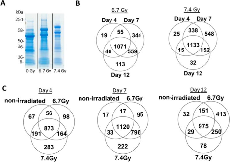
Figure 1.
Quantitative proteomic analysis of urine from non-human primates following gamma-irradiation reveals changes in the urine proteome. (A) Urine proteins from non-human primates (days 4, 7 and 12 post-exposure with 0, 6.7 or 7.4 Gy gamma-irradiation) were resolved by SDS-PAGE and visualized by Coomassie-staining. The gel image shows representative samples from non-human primates at 0Gy, 6.7 Gy and 7.4 Gy on day 4 post-irradiation. B) Venn diagram showing the number of total identified proteins that overlap between day 4, 7, and 12 post-irradiation following 6.7 Gy and 7.4 Gy exposures. C) Venn diagram showing total identified proteins from each time point (day 4, 7, 12) for 0 Gy, 6.7 Gy, and 7.4 Gy exposures.

