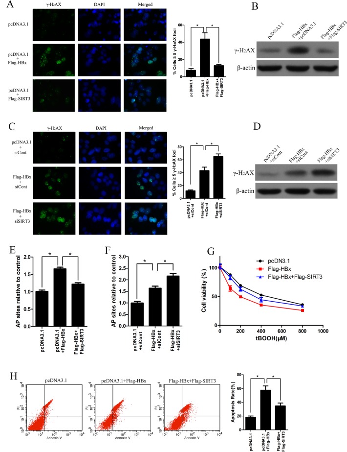
Fig 4. SIRT3 increases resistance of HBx-overexpressing cell to oxidant damage.
(A) Immunofluorescence of histone H2AX phosphorylation (γH2AX) in Huh-7 cells transfected with indicated plasmids for 3 days. Nuclei were stained with DAPI (left panel). Quantification of γH2AX foci (right panel). *, p<0.01. (B) Western blot analysis of γH2AX in Huh-7 cells transfected with indicated plasmids for 3 days. β-actin was used as a loading control. (C) Immunofluorescence of γH2AX in Huh-7 cells transfected with indicated plasmids and siRNA for 3 days. Nuclei were stained with DAPI (left panel). Quantification of γH2AX foci (right panel). *, p<0.05. (D) Western blot analysis of γH2AX in Huh-7 cells transfected with indicated plasmids and siRNA for 3 days. β-actin was used as a loading control. (E) AP sites numbers in Huh-7 cells transfected with indicated plasmids for 3 d were estimated by DNA Damage Quantification Kit, *, p<0.001. (F) AP sites numbers in Huh-7 cells transfected with indicated plasmids and siRNA for 3 d were estimated by DNA Damage Quantification Kit, *, p<0.01. (G) The cell viability in Huh-7 cells transfected with indicated plasmids under tBOOH treatment were measured by MTS assay. (H) Cell apoptosis in Huh-7 cells transfected with indicated plasmids under 100 μM tBOOH treated were measured by flow cytometry with Annexin V/PI. *, p<0.05.

