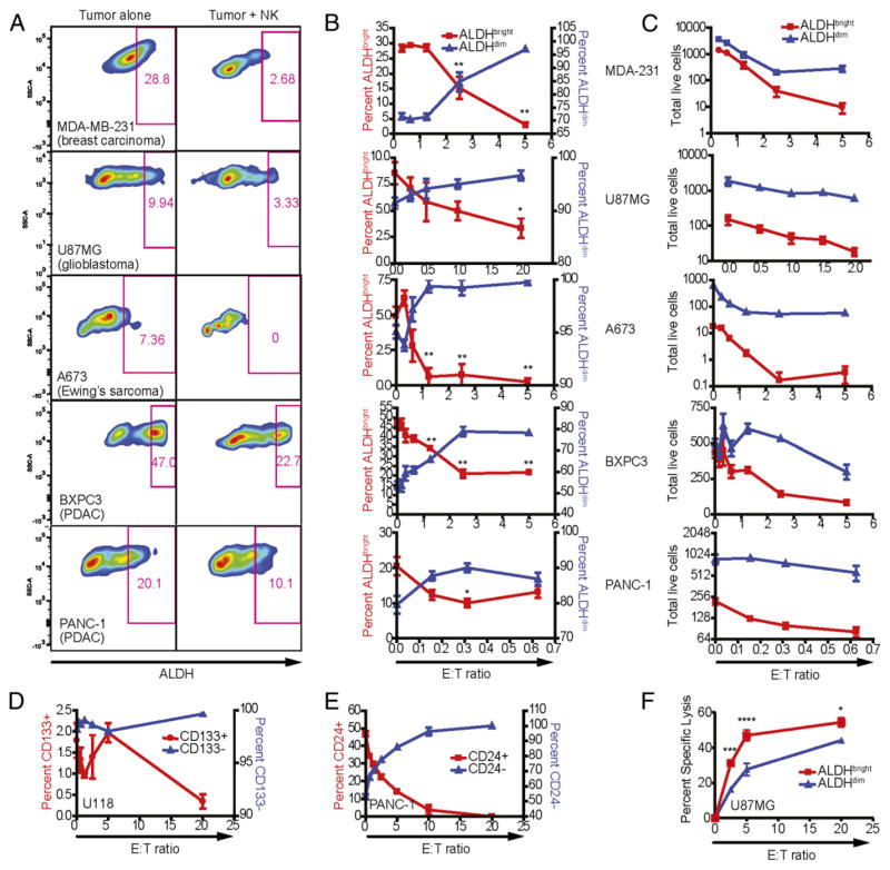
FIGURE 1.
Sensitivity of human tumor cell lines to NK cell lysis. Human NK cells were activated with IL-2, expanded for 2 wk, and cocultured 16–18 h with MDA-MB-231 (breast adenocarcinoma), U87MG (glioblastoma), A673 (Ewing’s sarcoma), BxPC3 (pancreatic ductal adenocarcinoma), and PANC-1 (pancreatic ductal adenocarcinoma) in the presence of 500 IU/ml rhIL-2. Phenotypes of remaining tumor cells were assessed by excluding 7-AAD+ events. (A) ALDH expression was assessed using the Aldefluor system in cultures with tumor cells alone and with NK cells at E:T ratios between 1:1 and 5:1. (B) The percentage of ALDHbright events on tumor cells was plotted (red; left y-axis) against the ALDHdim population (blue; right y-axis) at the indicated E:T ratios. (C) The total number of acquired events from (B) were plotted over the indicated E:T ratios. Reductions in ALDHbright populations in comparison with untreated cells were determined via one-way ANOVA with a Tukey posttest. *p < 0.05; **p < 0.01. (D) CSCs from the U118 glioblastoma were identified based on expression of surface marker CD133 (glioblastoma) following culture with NK cells at the specified E:T ratios. (E) Changes in the proportions of CD24-expressing and nonexpressing PANC-1 cells following NK coculture. (F) U87-MG cells were sorted for ALDH expression and then assessed for sensitivity to NK cell cytotoxicity in a 4-h 51Cr-release assay.

