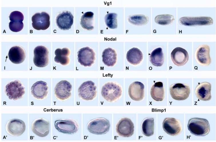
Fig. 1.
Expression of Vg1, Nodal, Lefty, Cerberus and Blimp1 in normal amphioxus embryos. Animal pole or anterior to left except as noted. Arrowheads = anterior limits of expression. (A-H) Vg1. (A) 2-cell stage. (B) 4-cell stage; dorsal view. (C) mid-blastula. (D) mid-gastrula; side view, blastopore at right. (E) Dorsal view of mid-gastrula in D. (F) Mid-neurula (15 hrs); side view. (G) Dorsal view of embryo in F. Expression in 4 left anterior somites; blastopore at right. (H) Late neurula (20 hrs); dorsal view; expression in left somites. (I-Q). Nodal. (I) Fertilized egg. Arrow shows second polar body. (J) 2-cell stage. (K) 8-cell stage. (L) Mid-blastula. (M) Surface view of blastula in K. (N) Late blastula; optical cross section. (O) Mid-gastrula. Side view; blastopore at right. (P). Blastopore view of the gastrula in O. (Q) Dorsal view of the gastrula in O. (R-Z) Lefty. (R) Very early blastula. (S) early-mid blastula. (T) Mid-blastula. (U) surface view of the embryo in T. (V) Vegetal pole view of the embryo in V. (W) Very early gastrula. (X) Mid-gastrula. (Y) Blastoporal view of late gastrula. (Z) Dorsal view of late gastrula. (A’-D’). Cerberus. (A’) Mid-gastrula; optical cross-section; animal pole at top. (B’) Mid-gastrula; blastopore at right. (C’) Early neurula; side view and (D’) dorsal view. Cerberus expression in anterior right paraxial mesoderm. (E’-H’) Blimp1. (E’) Mid-blastula. (F’). Mid-gastrula. Blastopore at right. (G’) Late gastrula. (H’) Early neurula.

