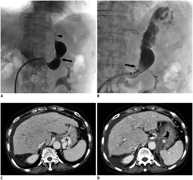
Fig. 1. 72-year-old female with GV and GRS.
A. 9 Fr TIPS sheath is placed in left renal vein and tip is in orifice to GRS. Contrast injection reveals GRS with waist (black arrow). Small inferior phrenic collateral vein (black arrowhead) is also seen. B. Vascular plug (arrow) is placed at waist of GRS. With gelfoam slurry injection, inferior phrenic collateral vein is spontaneously occluded and GV is completely filled with gelfoam mixture. C. Pre-procedural CT scan shows GV (asterisk). D. Complete thrombosis (asterisk) of GV is reported on post-procedural CT scan 4 days after PARTO. CT = computed tomography, GRS = gastrorenal shunt, GV = gastric varix, PARTO = plug-assisted retrograde transvenous obliteration, TIPS = transjugular intrahepatic portosystemic shunt

