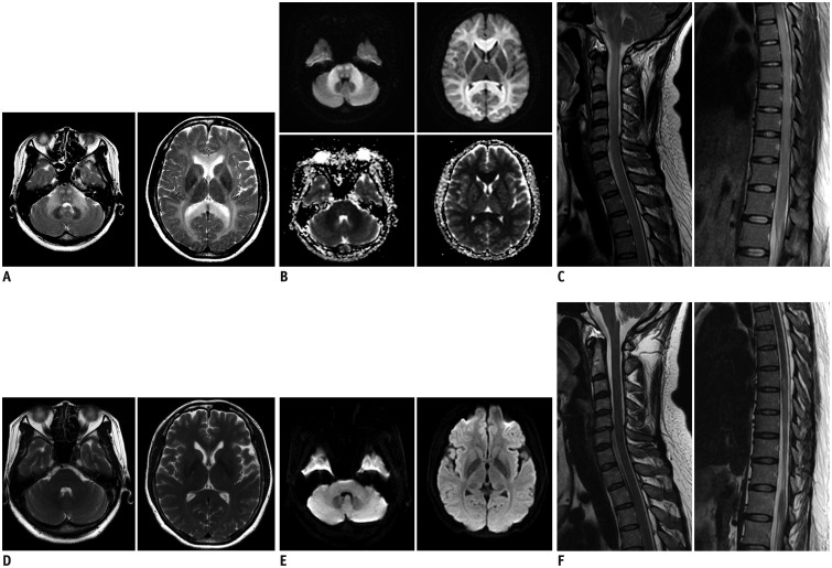
Fig. 1. Chlorfenapyr-induced toxic leukoencephalopathy in 44-year-old female patient.
A. Axial T2-weighted images show diffuse, bilaterally-symmetrical, and increased signal intensities involving periventricular white matter, corpus callosum, internal capsule, cerebellar white matter, and pons (corticospinal tract and central tegmental tract). B. Lesions show significant diffusion restriction on axial diffusion-weighted images and apparent diffusion coefficient map. C. Sagittal T2-weighted spinal images show diffuse enlargement of whole spinal cord with increased signal intensity. D. Follow-up axial T2-weighted images at 71 days after initial presentation show complete improvement in abnormal signal intensity within white matter throughout brain. E. Follow-up diffusion-weighted images confirmed complete resolution of previously noted restricted diffusion. F. Follow-up sagittal T2-weighted image of cervicothoracic spine shows improved general enlargement and hyperintensity of cervical and upper thoracic spinal cord. However, sagittal T2-weighted image of thoracolumbar spine shows residual hyperintensity in spinal cord below T7, with atrophic change.

